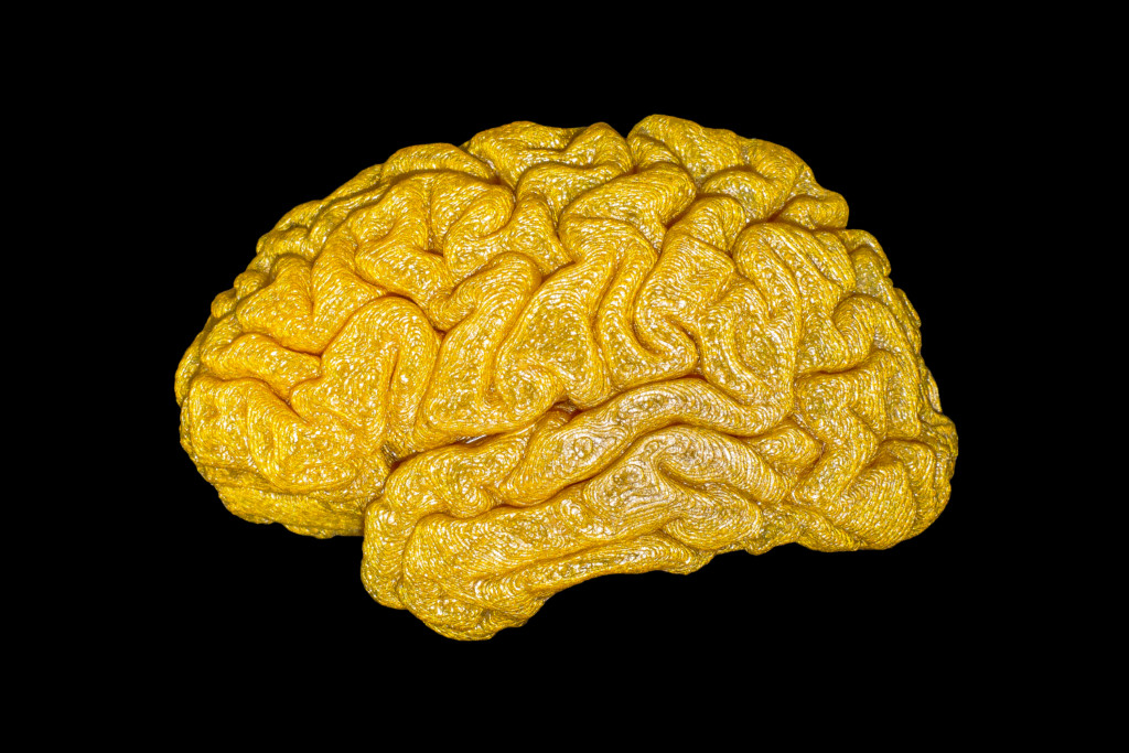
The Gold Standard: The 3D Brain
Photograph of 3D-printed reconstruction of the brain surface of a healthy female adult human brain. Neuroimaging sciences has seen the development of many novel techniques to investigate the structure and function of the living human brain. However, the gold standard is still considered to be post-mortem examinations and histological staining owing to the superior resolution and minimal source of artefacts. With the advent of advanced cutting-edge methodologies, which enable scientists to visualise the brain anatomy with a similar resolution to post mortem specimens, it might be high time we reconsidered what we define as our gold standard.
To create this work a colleague volunteered herself for a magnetic resonance imaging (MRI) session to acquire the original data which are contained in her T1-weighted scan (resolution of 0.9*0.9*0.9). These data were converted into 3D renders using the neuroimaging software package BrainVisa, and then exported to a printable format using Mashlab. The 3D print is made from golden Poly Lactic Acid filament with a 10% infill, a layer height of 0.1mm, and measures 4 x 6.5 x 3 cm. Picture was taking with an Apple iPhone 5 back camera 4.12mm f/2.4.
The Neuro Bureau
neuro-collaboration in action

